The Stages of Human Embryonic Development
Stage 3 begins when the blastocystic cavity first appears in the morula and ends when the zona (capsula) pellucida is shed as the embryo makes contact with the endometrial lining of the uterus. Stage 3 embryos are called free blastocysts. They have an estimated postfertilization age in vivo of between four and five days and measure 0.1 to 0.2 mm in diameter.
During Stage 3 the embryo separates into outer trophoblast cells and inner embryoblasts (inner cell mass) that will develop into the embryo proper. The site of the inner cell mass determines the embryonic pole of the blastocyst. The opposite side of the blastocyst becomes the abembryonic pole. Two axes can be distinguished; the polar (anterior-posterior) axis through the polar body and a perpendicular (dorso-ventral) axis at a right angle to the polar axis.
In vitro studies have shown that Stage 3 embryos go through four characteristic processes: 1) cavitation, 2) collapse/ expansion 3) hatching and 4) discarded cells.
The initiation of cavitation in the morula heralds the beginning of Stage 3. Cavitation occurs in vitro between post insemination days 5 and 7. Cavitation results from the accumulation of fluid inbetween the blastomeres. Since the total volume of the embryo does not change substantially during cavity formation, initial fluid accumulation appears to be a metabolic product of the blastomeres rather than transport from the outside.
Stage 3 blastocysts in vitro typically undergo repeated collapse and expansion before they escape from the zona pellucida. Collapse is rapid, occurring in less than five minutes whereas complete re-expansion requires several hours. The phenomenon has been called blastocyst “breathing” and occurs just before hatching. It is not known whether or not it occurs in vivo .
Hatching is the process by which the expanded blastocyst breaks through and escapes from the zona pellucida. It must occur before implantation into the endometrium is possible. Hatching of healthy blastocysts in vitro usually occurs between post insemination days 7 and 8. After hatching the blastocyst enlarges substantially, the trophoblast cells thin and tight junctions form between them, and , small fimbriated trophoblast projections appear in the region of the blastocyst where attachment usually occurs.
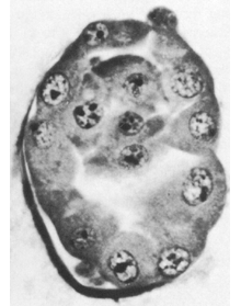
Examination of in vitro Stage 3 blastocysts with TEM reveals two types of cells that are apparently discarded by the developing embryo, 1) sequestered cells and 2) isolated cells. Sequestered cells are groups of cells located between the zona pellucida and the trophoblast, while iIsolated cells are found mainly in the blastocystic cavity.
| Browse the section images |
| 3D reconstructions |
| More info ... |