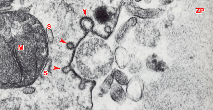
Fig. 14. Very high power TEM of the cell membrane of a stage 1a embryo in vitro, 24 hours after insemination (original magnification x70,000). Notice simultaneous exocytosis of a cortical granule and formation of micropinocytotic caveolae (arrowheads). The cell membrane of the embryo is dense and has a striated appearance in this region. A mitochondrion (M) is closely associated with elements of the smooth endoplasmic reticulum (S).
ZP, zona pellucida.
(From: Sathananthan and Trounson, 1982. Reproduced with permission of the publisher, John Wiley & Sons.)
|
|