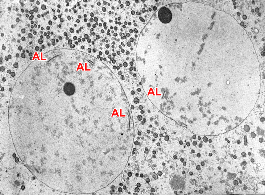
Fig. 15. TEM of a stage 1b embryo in vivo near the end of the pronuclear period when the pronuclei lie in close proximity. The embryo was flushed from the uterine tube 26 hours after reported copulation and at an unknown interval after ovulation. The pronuclear plasm is highly hydrated and consists of finely dispersed granular material. The number of nucleoli increases with age. A few annulate lamellae (AL) are evident inside the pronuclei.
(From: Zamboni, 1971. Reproduced with permission of the publisher, Lippincott, Williams, & Wilkins.)
 |