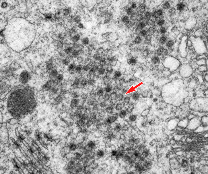
Fig. 18. High power TEM of the cytoplasm of a stage 1b embryo in vitro, 24 hours post-insemination (original magnification x19,700). The section is tangential to the surface of annulate lamellae showing the ring-shaped structure of the pores and their hexagonal arrangement (arrow).
(From: Soupart and Strong, 1974. Reproduced with permission of the American Society for Reproductive Medicine.)
|
|