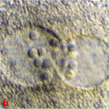
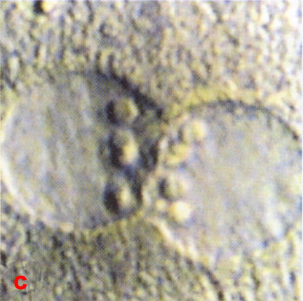
Fig. 6. LMs of a series of close-up views of pronuclei in stage 1b embryos in vitro showing nucleolar distribution.
(From: Veeck and Zaninovic´, 2003. Reproduced with permission of the publisher, Taylor & Francis Group.)
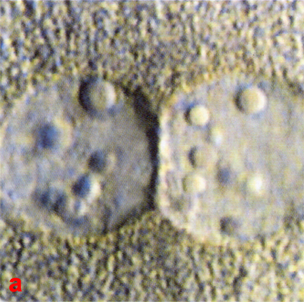
a) The nucleoli are scattered in both pronuclei.
b) The nucleoli are aligned in the pronucleus on the right but scattered in the pronucleus on the left.
c) Both pronuclei have aligned nucleoli.