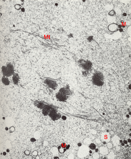
Fig. 7 EM of a stage 1c embryo in vitro, during the first cleavage, 40 hours post-insemination (original magnification x14,580). The mitotic spindle consists of microtubules (Mt) somewhat disoriented and ending abruptly in the cytoplasm at either pole. No centriole or asters are evident. Three sets of chromosomes are at the equator of the spindle with one already moved toward a pole. The chromation appears to be separating into two chromosomes, signifying the beginning of anaphase. The vesicular smooth endoplasmic reticulum (S) is very dilated. Unusual C-shaped mitochondria (M) are present in the cytoplasm.
(From: Sathananthan and Trounson, 1985. Reproduced with permission of the publisher, John Wiley & Sons.)
|
|