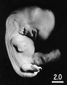
Lateral view of embryo No. 6520 after fixation.
O'Rahilly and Müller (1987) Fig. 17-2
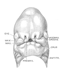
Intermediate development of the nose and future upper jaw.
O'Rahilly and Müller (1987) Fig. 17-3
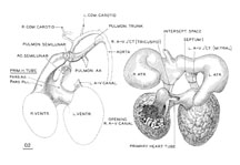
Reconstruction of the endocardium of the heart and its arterial trunks in embryo No. 6520.
O'Rahilly and Müller (1987) Fig. 17-7
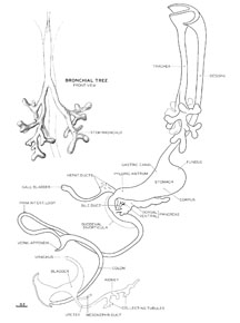
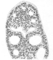
Reconstruction of the epithelial core of the alimentary canal of embryo No. 6520, typical of stage 17 embryos.
O'Rahilly and Müller (1987) Fig. 17-8
Cross section of the duodenal epithelium.
O'Rahilly and Müller (1987) Fig. 17-9
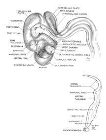
Reconstruction of the right half of the brain.
O'Rahilly and Müller (1987) Fig.17-12
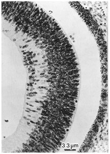
Photomicrograph of the eye.
O'Rahilly and Müller (1987) Fig. 17-13
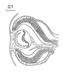
Diagram of cross sectioned eye.
O'Rahilly and Müller (1987) Fig. 17-14