Index of Stage 2 In Vitro Light Micrographs
(1 of 2)
Click on an image to see details.
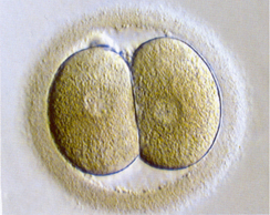
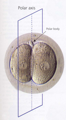
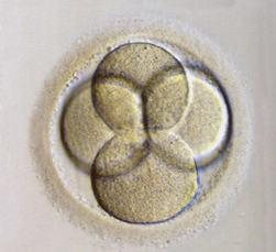
Fig. 1. Embryo after first cleavage is completed
Fig. 2. Polar axis
Fig. 3. 4-cell embryo
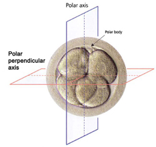
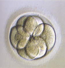
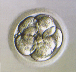
Fig. 4. Axes at 4 cells
Fig. 5. 6-cell embryo
Fig. 6. 8-cell embryo
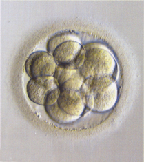
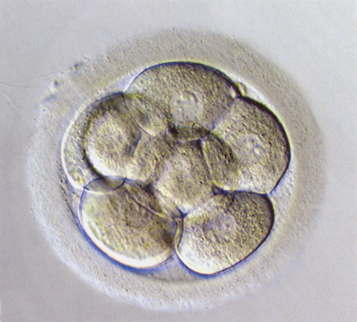
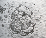
Fig. 7. 12-cell embryo
Fig. 8. Initial stage of compaction
Fig. 9. Morula at the beginning of compaction