Index of Stage 2 In Vitro Scanning Electron Micrographs
(1 of 2)
Click on an image to see details.
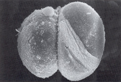
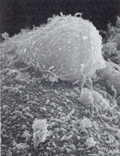
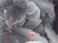
Fig. 16. 2-cell embryo
Fig. 17. Close up of second polar body seen in the Figure 16 specimen
Fig. 18. Periphery of a 5-cell embryo
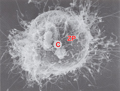
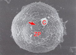
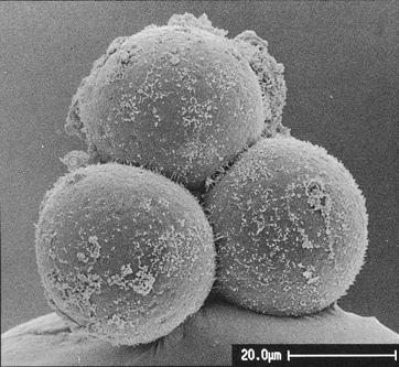
Fig. 19. Outer surface of the zona pellucida
Fig. 20. One corona cell attached to the zona pellucida
Fig. 21. 4-cell embryo