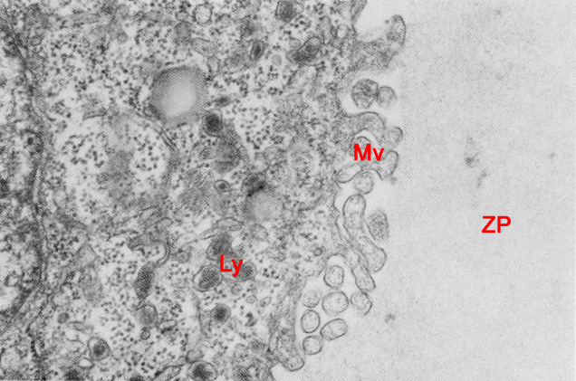
Fig. 15. Very high power TEM of a portion of the trophoblast/zona pellucida junction area of a stage 3 embryo in vitro, 145-170 hours post-insemination (original magnification x34,200). The plasma membrane specializations in the trophoblast distinguish it from the inner cell mass. Highly developed microvilli and complex intercellular junctions give the trophoblast the appearance of specialized epithelium. Microvilli are most numerous on the outer surface of the trophoblast where they project toward the zona pellucida.
Mv, microvilli; Ly, lysosome; ZP, zona pellucida.
(From: Lopata, Kohlman, and Kellow, 1982. Reproduced with permission of the publisher, John Wiley & Sons.)
|