Index of Electron Micrographs of Stage 4 Human Embryos In Vitro
(2 of 3)
Click on an image to see details.
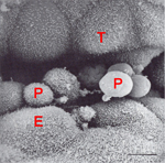
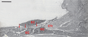
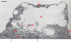
Fig. 7. Contact area between the embryo and the endometrial cell culture
Fig. 8. Details of the contact zone between the endometrial cells and the cytotrophoblast.
Fig. 9.Blastocyst attached to the endometrial cell culture.
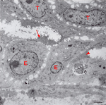
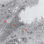
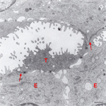
Fig. 10. The most peripheral contact area between two trophoblast cells and cultured endometrial cells.
Fig. 11. Contact area showing apical junctional complexes and desmosomes between penetrating trophoblast and endometrial epithelial cells.
Fig. 12. Contact area showing apical junctional complexes and desmosomes between the endometrial epithelium and a trophoblast cell.