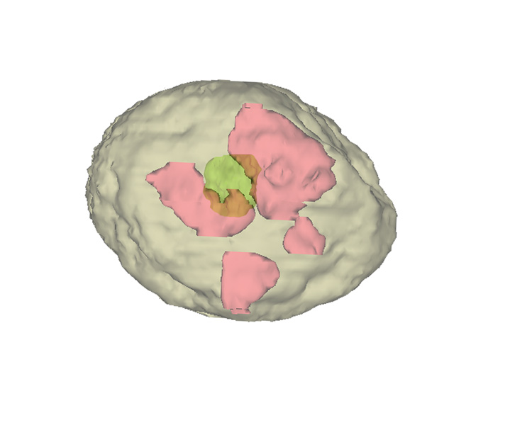
Computer 3D reconstruction of the attachment area (pink)
. In these areas the trophoblastic syncytium is fusing with the endometrial epithelium. The inner cell mass (green) is visible through the semitransparent blastocyst wall.
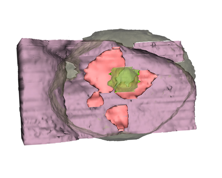
View of the blastocyst from the uterine wall, highlighting the attachment sites. The endometrial epithelium has been omitted.
To compare plate 5 from Heuser and Streeter, (1941) with our 3D-reconstructions, click here
To turn off the overlay click here
View of the attaching blastocyst looking towards the uterine wall. The blastocyst wall has been made transparent to show the attachment sites and the inner cell mass
 |
 |
|
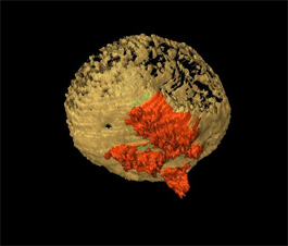
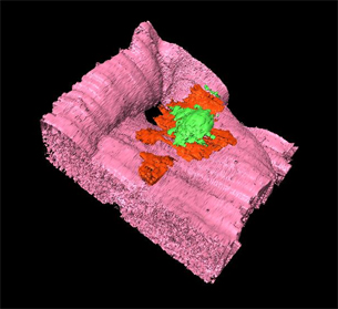
| Volumetric reconstruction of the blastocyst showing the attachment sites in red. Click on the image to view the full animation. | Volumetric reconstruction of the uterine wall at the attachment site. The inner cell mass (green) is also shown. Click on the image to view the full animation |