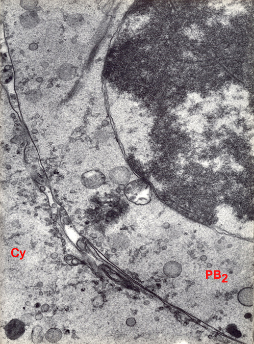
Fig. 12. TEM of a stage 1a embryo in vivo showing the junction area between the embryo and the second polar body, 26 hours post-copulation. The embryo was flushed from the uterine tube at an unknown interval after ovulation. The chromosomes of the second polar body (PB2) are surrounded by a double envelope similar to a nucleus. This feature and the absence of cortical granules distinguishes the second polar body from the first.
Cy, cytoplasm.
(From: Zamboni, et al., 1966. Reproduced with permission of the publisher, Rockerfeller University Press.)
 |