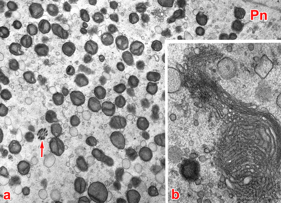
Fig. 20. High power TEM of a stage 1b embryo in vivo showing cytoplasmic organelles, 26 hours post-copulation. The embryo was flushed from the uterine tube at an unknown interval after ovulation.
(From: Zamboni, 1971. Reproduced with permission of the publisher, Lippiocott Williams &Wilkins.)
a) Mitochondria are numerous and uniformly distributed throughout the cytoplasm. Many are in close association with smooth endoplasmic reticulum vesicles. The naked fibers of the flagellum of the fertilizing sperm (arrow) are relatively close to one pronucleus (Pn) that is likely the paternal (male) one. No cortical granules are present.
b) Unique to the stage 1b embryo is the formation of prominent Golgi complexes in the central cytoplasm in close association with the pronuclei. The complexes consist of numerous small vesicles and flattened cisternae in parallel arrangement. They progressively increase in number to reach a maximum shortly before the first cleavage.
 |
|