Index of Stage 2 In Vivo and In Vitro Transmission Electron Micrographs
(1 of 4)
Click on an image to see details.
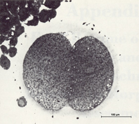
Fig. 27. 2-cell embryo undergoing mitosis
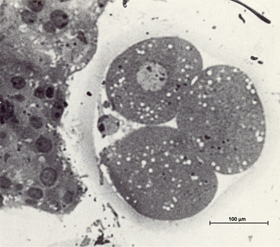
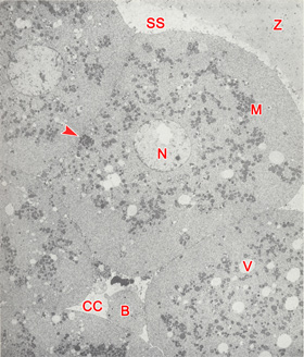
Fig. 28. 3-cell embryo
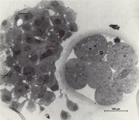
Fig. 29. 8-cell embryo
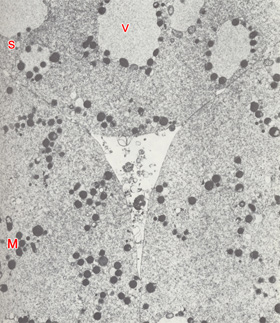
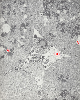
Fig. 30. Central cavity of a 4-cell embryo
Fig. 31. Central cavity of an 8-cell embryo
Fig. 32. Central cavity of an 8-cell embryo
Fig. 33. 7-cell embryo
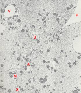
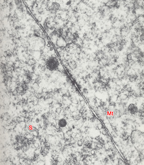
Fig. 34. Cytoplasm and cell junction of a 2-cell embryo
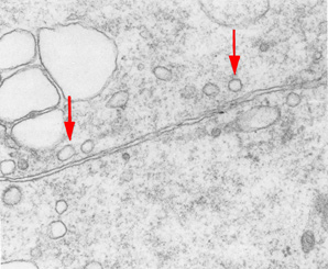
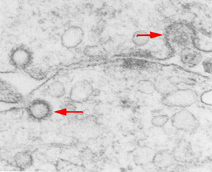
Fig. 35. Cell contact area of an embryo
Fig. 36. Cell contact area in a 4-cell embryo
Fig. 37. Cell junction of a 4-cell embryo
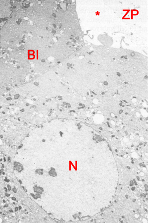
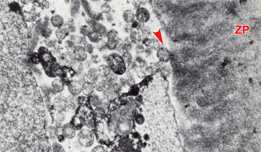
Fig. 38. Subzonal space of an 8-cell embryo
