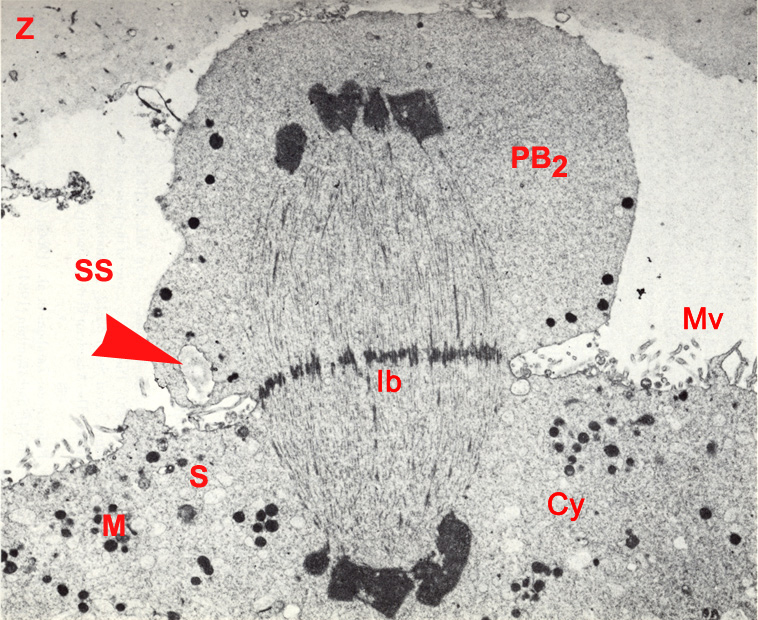
Fig. 8. TEM of a stage 1a embryo in vitro showing telophase II and expulsion of the second polar body, 3 hours post-insemination (original magnification x8,620). The zona pellucida (Z) is pushed outward thereby enlarging the subzonal space (SS) around the second polar body (PB2). The second meiotic spindle is barrel-shaped and composed of numerouse microtubules. At the two poles of the spindle are chromatids that have separated and are beginning to organize into distinct nuclei. Centrioles (asters) are absent. The microtubules extend to the chromatin masses. A pocket of zona pellucida material (arrowhead) is visible in the second polar body. The cytoplasmic constriction is deep on either side of the dense interbody (Ib) which has contracted.
M, mitochondria; Mv, microvilli; Cy, cytoplasm; S, vesicular smooth endoplasmic reticulum.
(From: Sathananthan, Trounson, and Wood, 1986. Reproduced with permission of the publisher, Greenwood Publishing Group.)
|