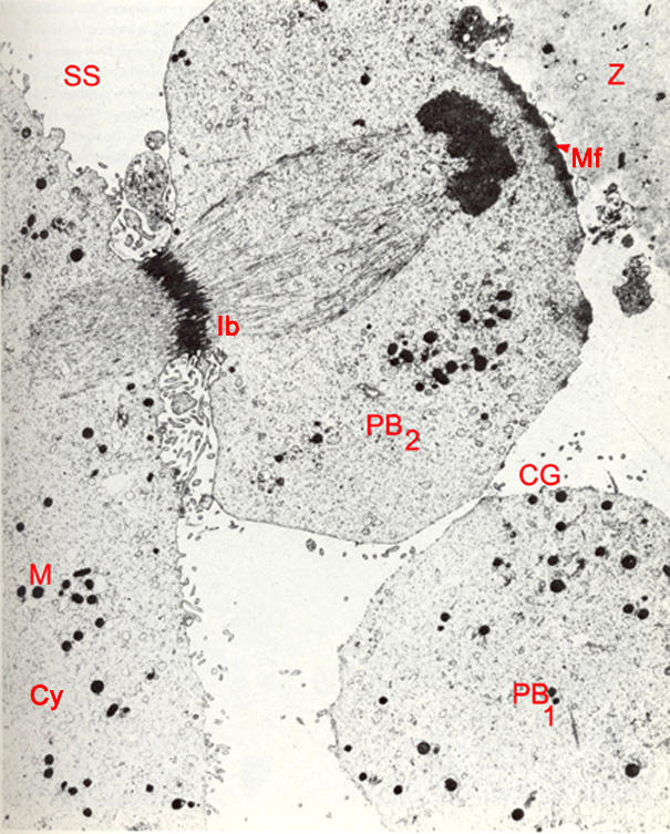
Fig. 9 TEM of a stage 1a embryo in vitro showing the abstriction of the second polar body, 3 hours post-insemination (original magnification x6,810). The spindle is sectioned obliquely and has lost its barrel shape. Cortical granules have released their contents around the embryo suface but a few are intact in the second polar body. The cytoplasmic furrow has deepened, constricting the dense interbody region (Ib) of the spindle. The spindle connects the second polar body (PB2) to the cytoplasm (Cy) by a narrow bridge. The chromatin masses in the second polar body have fused and are organizing into a nucleus. The advancing edge of the second polar body exhibits a band of minute microfilaments (Mf) deep to its membrane which are seen here in cross section. The microfilaments are thought to play a role in polar body constriction. The embryo and second polar body are usually devoid of cortical granules (CG). Numerous microvilli and cytoplasmic debris are located in the furrow between the embryo and second polar body. They arise mainly from the embryo. The second polar body has a smooth contour.
M, dense mitochondria; SS, subzonal space; PB1, first polar body; Z, zona pellucida.
(From: Lopata et al, 1980 . Reproduced with permission of the American Society for Reproductive Medicine.)
|