Index of Stage 2 In Vivo and In Vitro Transmission Electron Micrographs
(2 of 4)
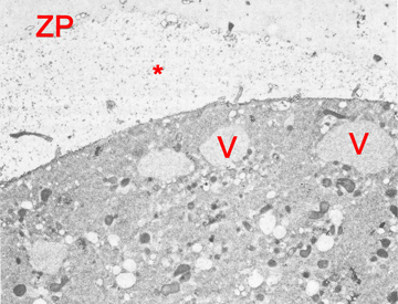
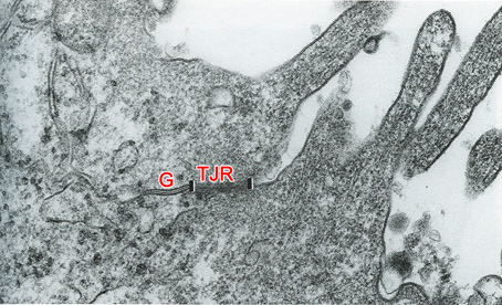
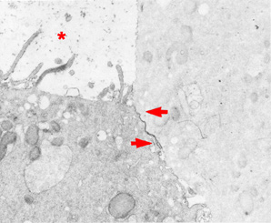
Fig. 39. Blastomere periphery of a 7-cell embryo
Fig. 40. Adjacent surfaces of two polar cells in a morula
Fig. 41. A primordial junctional complex near the subzonal space in a 7-cell embryo
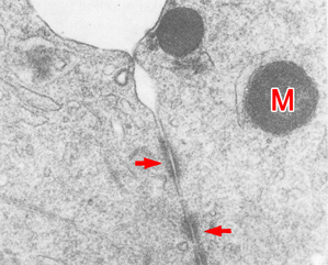
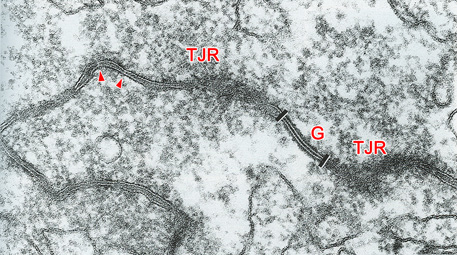
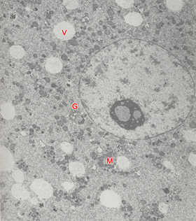
Fig. 42. Two rudimentary desmosomes in a 2-cell embryo
Fig. 43. Adjacent surfaces of a polar and an apolar cell in a morula
Fig. 44. Blastomere of an 8-cell embryo
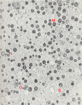
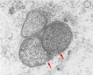
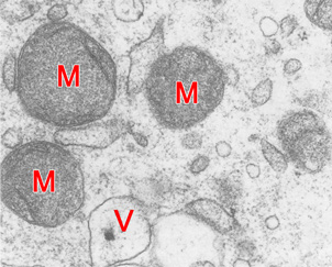
Fig. 45. Cytoplasmic organization in a blastomere of a 4-cell embryo
Fig. 46. Section of a blastomere of an embryo
Fig. 47. Mitochondria in an early stage 2 embryo
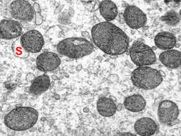
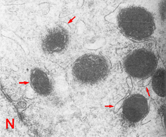
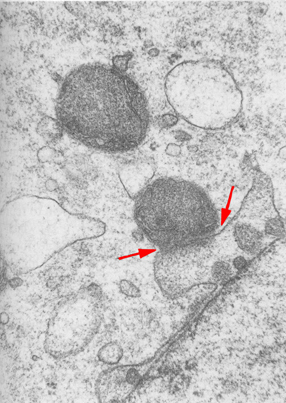
Fig. 48. Detail of mitochondria
Fig. 49. Flattened saccules associated with mitochondria in a 4-cell embryo
Fig. 50. Blebs protruding from the nuclear membrane associated with mitochondria in a 4-cell embryo