Index of Stage 2 In Vivo and In Vitro Transmission Electron Micrographs
(3 of 4)
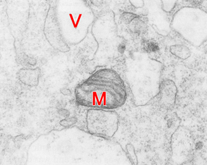
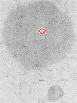
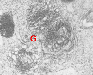
Fig. 51. Detail of mitochondrion in a 7-cell embryo
Fig. 52. Crystallin inclusions in the cytoplasm of a 16-cell embryo
Fig. 53. Well developed Golgi complex in the cytoplasm of a 4-cell embryo
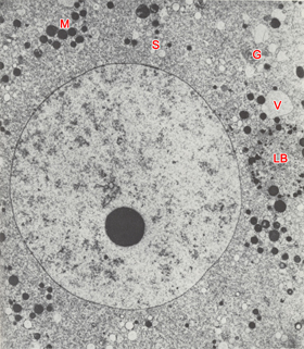
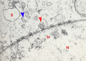

Fig. 54. Nucleus and surrounding cytoplasm of a 2-cell embryo
Fig. 55. Nuclear organization of a blastomere in a 4-cell embryo
Fig. 56. Blebbing of the nuclear membrane in a blastomere of 4-cell embryo
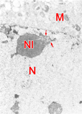
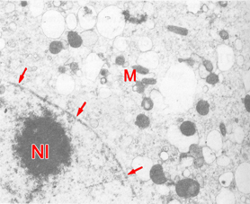
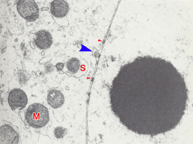
Fig. 57. Beginning of differentiation of the nucleolus in a 7-cell embryo
Fig. 58. Blebbing absence of the nuclear membrane of a 16-cell embryo
Fig. 59. Nuclear organization of a blastomere in a 4-cell embryo
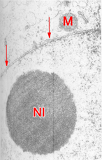
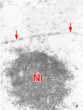
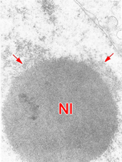
Fig. 60. Nuclear membrane and nucleolus of a 2-cell embryo
Fig. 61. Blebbing absence of the nuclear membrane of a 16-cell embryo
Fig. 62. Nucleolus of a blastomere of a 4-cell embryo