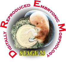
Stage 23 embryos have a greatest length of 23 to 32.2 mm and an estimated postfertilization age of 53 to 58 days. Over 90% of the more than 4,500 named structures in the adult body are present. All named muscles can be identified. The head is larger and more rounded than in previous stages, and the subcutaneous venous plexus lies close to the vertex. The limbs are more elongated and the hands and feet can overlap each other. The external genitalia are prominent but not yet sexually distinct. The insula and subdivisions of the lateral ventricle can be identified in the cerebral vesicle (hemisphere), the superior and inferior colliculi are evident in the tectum of the mesencephalon and the external granular layer of the cerebellum has formed in the alar plate of the metencephalon. Many nuclei, fiber tracts, decussations and commissures are evident in the brainstem and all four parasympathetic ganglia in the head are present. The upper and lower eyelids are beginning to fuse with each other both laterally and medially. The spiral cochlear duct attains its definitive arrangement of 2 ½ turns. The laryngeal cartilages are present, the palatine shelves are fusing with each other and the nasal septum, and the salivary gland ducts have begun secondary branching. Ossification has begun in the skull and the long bones. Most of the named arteries to the brain can be identified and the left superior vena cava is no longer present. The kidney exhibits 4 to 5 orders of tubules, and tubules can be identified in the testis but are not yet fused with the rete testis and mesonephric duct.
Stage 23 is represented in the DREM databases by Carnegie embryo # 9226 that had been given a grade of excellent. It has a greatest length of 31 mm after fixation and is considered to be advanced in the stage. The estimated post fertilization age is 56 days.
The embryo was prepared for microscopic examination in 1954. It was fixed in formol, embedded in celloidin and paraffin and serially sectioned transverse to the long axis at 12 microns. The sections were mounted on glass slides and stained with Azan. There are 2090 sections through the embryo, they are mounted on 209 slides .
The DREM database includes 209 of the 2090 sections. Approximately every 10th section was digitally restored and labeled, and can be viewed at four magnifications. The aligned sections were used to produce 3D reconstructions. Animations of the reconstructions and flythrough animations of the aligned sections are also included on the disks. For anyone who wishes to use them for other reconstructions, research or presentations, all the original section images are available as individual .jpg files. A self-extracting archive of the section images is also available.
Embryo # 9226 has been studied extensively and there are several publications dealing with various aspects of its morphology. A selection of figures from most of these articles is included on the disk.
Instructions for using the disks can be found by going to the Instructions section from the opening screen.


