Index of Photographs and Figures Relating to Embryo 9226 (1 of 4)
Click on an image to see details.
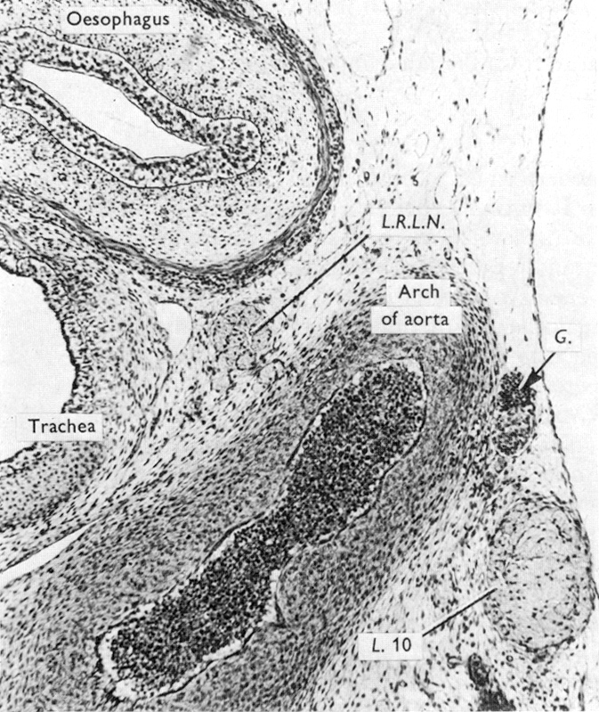
A
ganglion on a cardiac nerve at the left side of the arch of the aorta.
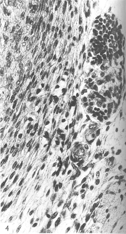
The ganglion of Figure 3 shown
at a higher magnification.
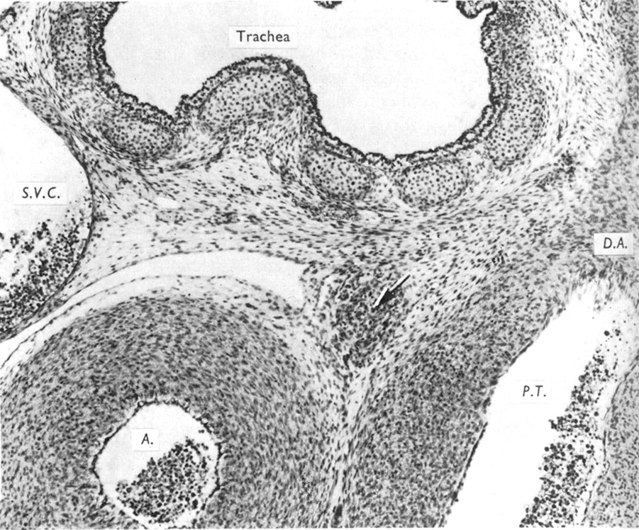
The caudal portion of the aorticopulmonary
ganglion (arrow).
Gardner and O'Rahilly (1976)
Fig. 3
Gardner and O'Rahilly
(1976) Fig. 4
Gardner and O'Rahilly (1976) Fig. 5
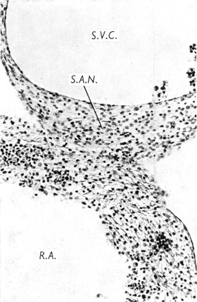
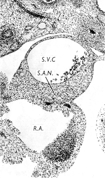
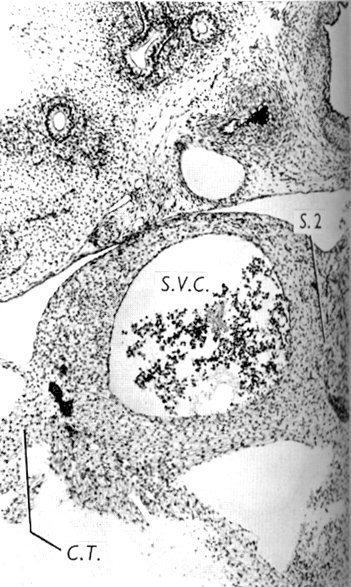
The sinu-atrial
node in the anterior wall of the superior vena cava.
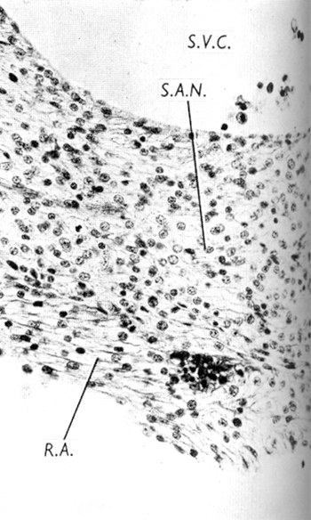
The sinu-atrial node somewhat
caudal to the level of Figure 7. Note its increased thickness.
The sinu-atrial node and wall
of right atrium of Figure 8.
Gardner and O'Rahilly (1976) Fig. 7
Gardner and O'Rahilly (1976) Fig. 8
Gardner and O'Rahilly (1976) Fig. 9
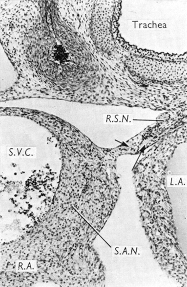
At a level caudal
to that of Figure 8.
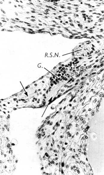
The venous mesocardium of Figure
10, with the right sinal nerve, ganglion cells and nerve filaments.
Nodal tissue completely encircles
the superior vena cava, but is thinning anteriorly.
Gardner and O'Rahilly (1976) Fig. 10
Gardner and O'Rahilly (1976) Fig. 11
Gardner and O'Rahilly (1976) Fig. 12