Index of Stage 3 Cavitation In Vitro
(3 of 3)
Click on an image to see details.
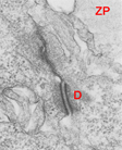
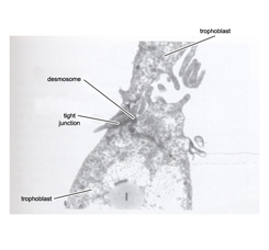
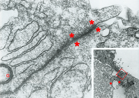
Fig. 19. Junctional complex between two trophoblast cells
Fig. 20. Cell junction between two trophoblast cells
Fig. 21. Apical tight junction in the appositional region of two trophoblast cells
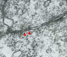
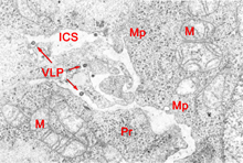
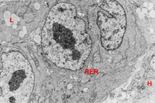
Fig. 22. Basal tight junction between two trophoblast cells
Fig. 23. Virus like particles in the intercellular space
Fig. 24. Epiblast/hypoblast junction

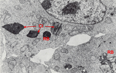
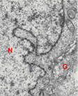
Fig. 25. Two adjacent epiblast cells of the inner cell mass
Fig. 26. Cytoplasmic inclusions in an epiblast cell
Fig. 27. Portion of the nucleus of an epiblast cell
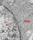
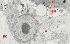
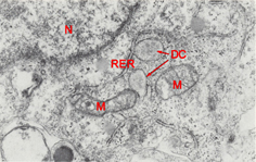
Fig. 28. Portion of the nucleus of an epiblast cell
Fig. 29. Hypoblast cell
Fig. 30. Hypoblast cell