Index of Stage 3 Cavitation In Vitro
(2 of 3)
Click on an image to see details.
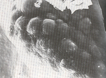
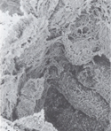
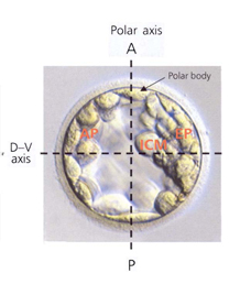
Fig. 10. Inner cell mass
Fig. 11. Inside view
Fig. 12. Axes of the blastocyst
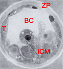
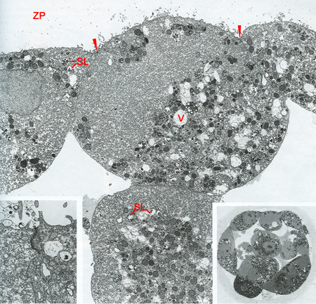
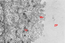
Fig. 13. Blastocystic cavity, inner cell mass, trophoblast, and zona pellucida
Fig. 14. Polar trophoblast
Fig. 15. Trophoblast/zona pellucida junction area
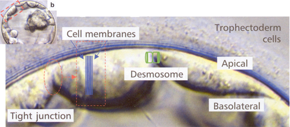
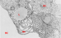
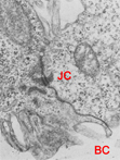
Fig. 16. Schematic of trophoblast cell junctions
Fig. 17. Junctional region of two trophoblast cells
Fig.18. Junctional complex between two trophoblast cells