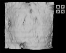
Click on an image to start the animation.
 |
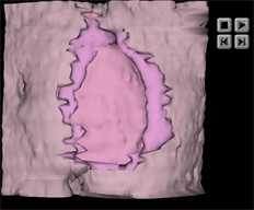 |
| Animation 1: Oscillation of the tissue block containing the implantation site surrounded by the mouths of several endometrial glands. The surface view fades to show the trophoblast embedded in the decidua. The edge of the implantation site is lined with disrupted endometrial epithelium. | Animation 2: Continuation of animation 1. The decidua fades to reveal the rotating trophoblast which then fades to show the embryonic mass comprising, epiblast (green), hypoblast (yellow) and amnioblast (white). |
These 3D animations were made from the WinSurf reconstructions, however, to enable users to better study the reconstructions, we are also providing the original WinSurf files together with a WinSurf Viewer. This viewer will allow users to open WinSurf data files and view reconstructions with all the display options of the full reconstruction program. Models can be rotated about all three orthogonal axes, users can zoom in and out when viewing models and models can be translated. Also, the color and opacity of different objects in the models can be varied.