| Embryo 8155 - Stage 5a 2 |
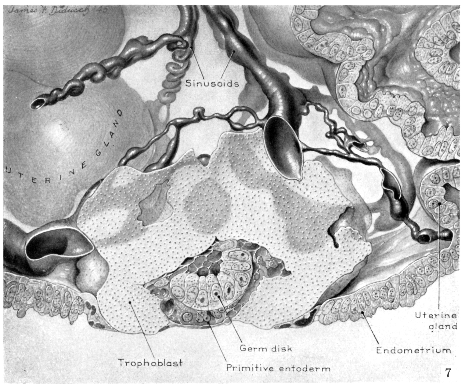
Fig. 7. Hertig and Rock, 1949. |
| A drawing of one-half the reconstructed ovum viewed at the level of the greatest diameter of the embryo. The top section of the reconstruction coincides with the photomicrograph shown in figure 3. The trophoblast is represented by the solid stippled part, whereas the essential histological details of the remainder of the ovum are shown. Note the damaged surface epithelium in juxtaposition to trophoblast. |
|
| Click Here or
on the figure to view a full size image |
|