Index of Stage 1b In Vitro
(1 of 3)
Click on an image to see details.
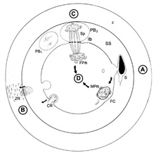
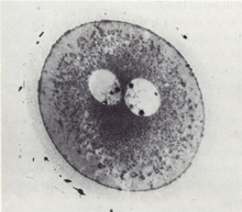
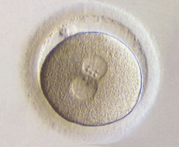
Fig. 1. Schematic of events of fertilizations of stage 1a and 1b embryos
Fig. 2. Narrow interpronuclear area
Fig. 3. Two pronuclei of approximate equal size
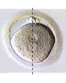
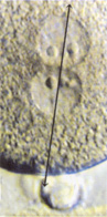
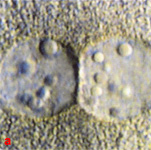
Fig. 4. Pronuclear alignment along the polar axis
Fig. 5. Close up view of polarized pronuclei
Fig. 6. A series of close up views of pronuclei showing nucleolar distribution
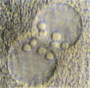
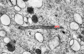
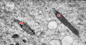
Fig. 7. Close up view showing good nucleoli distribution
Fig. 8. Longitudinal section through a wavy part of the fertilizing sperm flagellum
Fig. 9. High power TEM of the cytoplasm