Index of Published Figures Relating to Embryo 9226 (2 of 4)
Click on an image to see details.
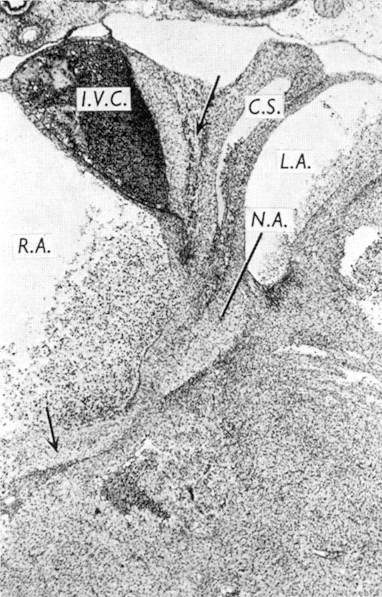
The caudal portion of the atrioventricular node, which contains a nodal artery.
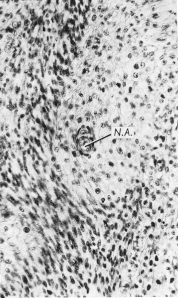
The most caudal portion of the atrioventricular node, with nodal artery.
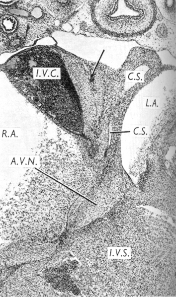
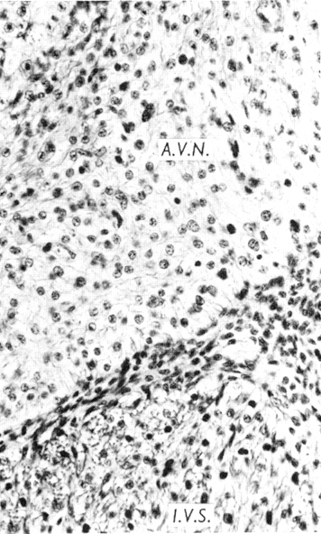
The atrioventricular node and interventricular septum at a more rostral level.
Gardner and O'Rahilly (1976) Fig. 15
Gardner and O'Rahilly (1976) Fig. 16
Gardner and O'Rahilly (1976) Fig. 17
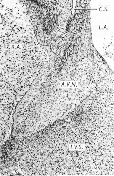
The atrioventricular node of Figure 17. Note sharp separation from interventricular septum.
The atrioventricular node of Figure 17 at a higher magnification.
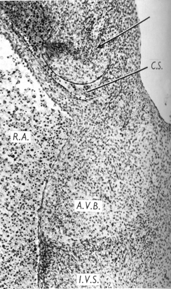
At the level of the opening of the coronary sinus the atrioventricular node is becoming the atrioventricular bundle.
Gardner and O'Rahilly (1976) Fig. 18
Gardner and O'Rahilly (1976) Fig. 19
Gardner and O' Rahilly (1976) Fig. 20
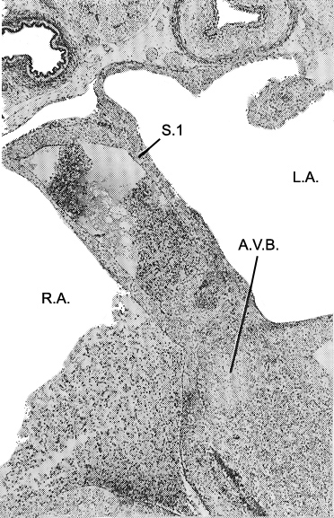
The septum primum and the atrioventricular bundle.
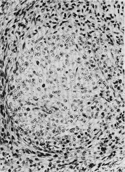
The atrioventricular bundle.
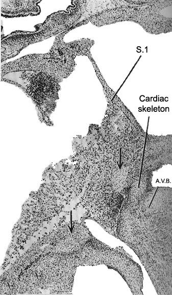
The atrioventricular bundle,
immediately behind which is a portion of the developing cardiac skeleton.
Gardner and O'Rahilly (1976) Fig. 21
Gardner and O'Rahilly (1976) Fig. 22
Gardner and O'Rahilly (1976) Fig. 23