Index of Published Figures Relating to Embryo 9226 (3 of 4)

The right limb of the atrioventricular bundle.
The right limb of Figure 24.
The right limb of Figure 24 at a higher magnification.
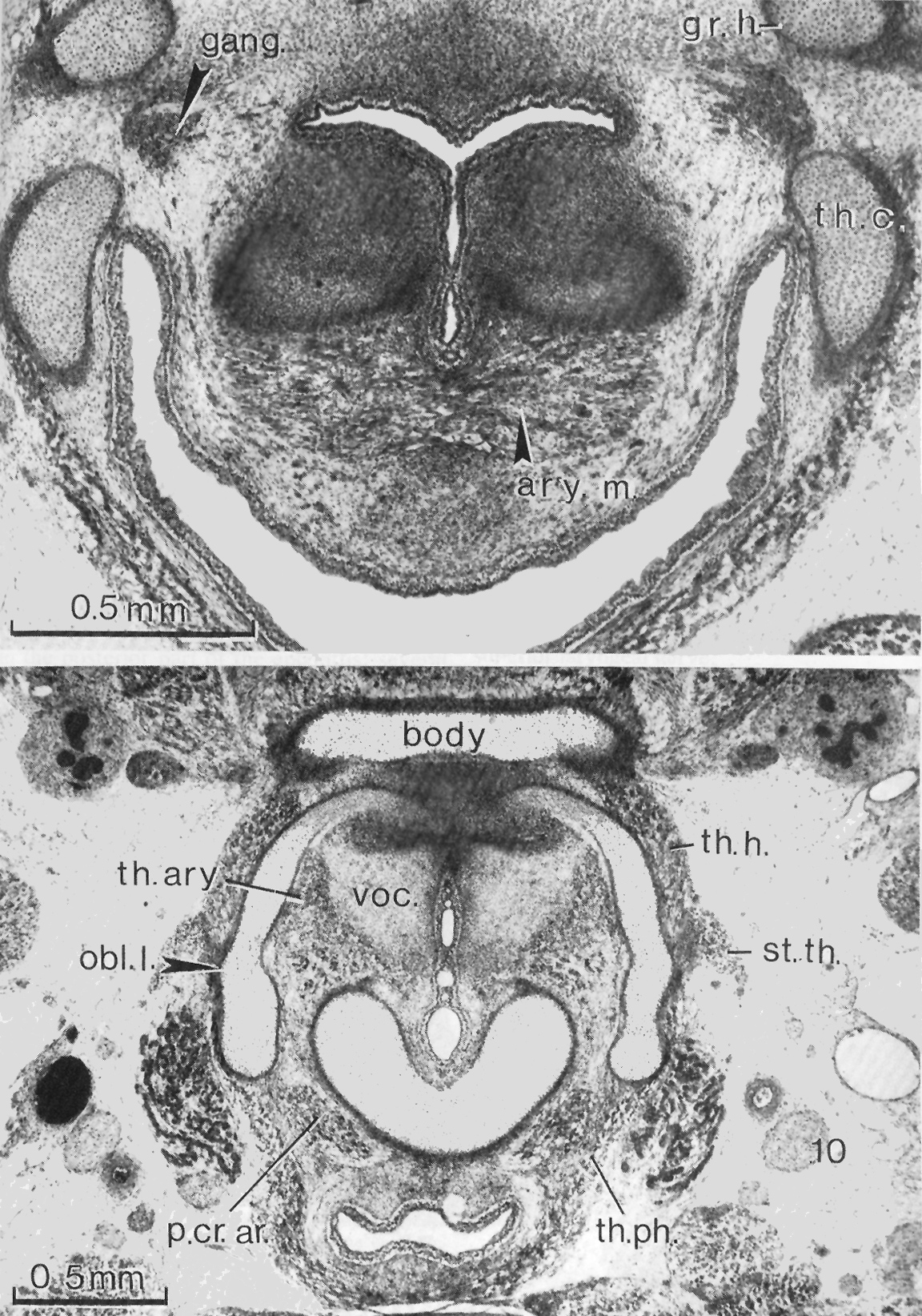
Transverse sections of No. 9226
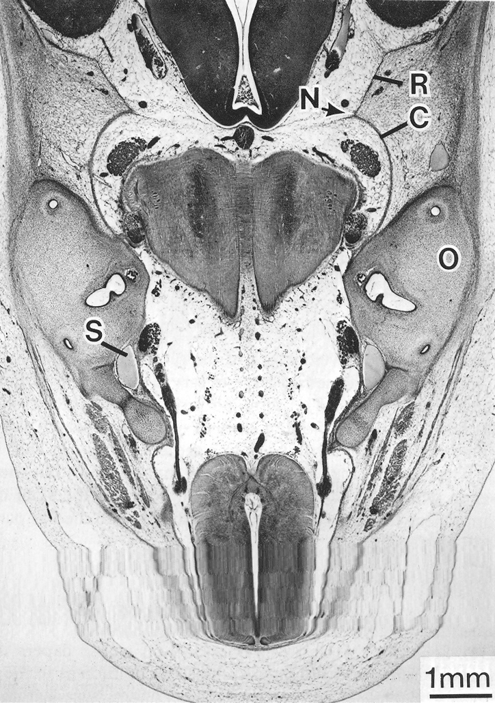
Transverse
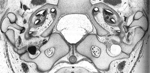
section through the base of the skull at 31 mm, showing the hypoglossal
canal in the occipital cartilage on each side.
In a, the greater horn
of the hyoid and the thyroid lamina can be seen on each side. In b,
the body of the hyoid, thyroid laminae, and cricoid cartilage are visible.
Gardner and O'Rahilly (1976)
Figs. 24-26
Müller, O'Rahilly and Tucker (1981)
Fig. 7
O'Rahilly, Müller and
Meyer (1983) Fig. 4a
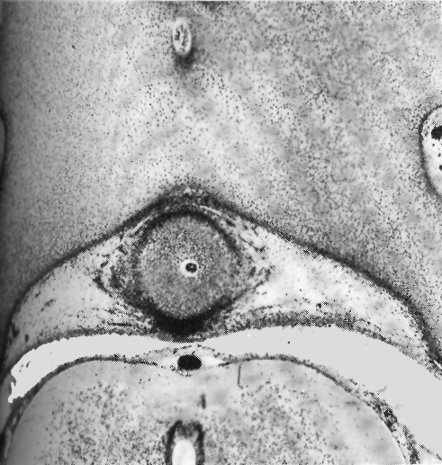
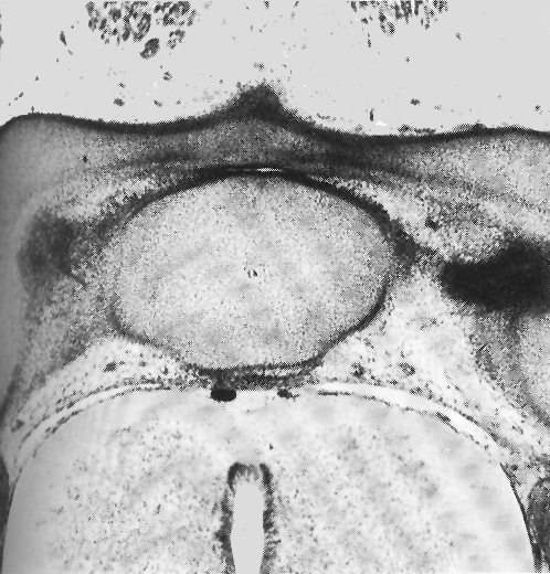
Occipito-axial joint at 31 mm, showing the beginning articular cavity.
Occipito-axial
joint at 31 mm, showing the beginning articular cavity.
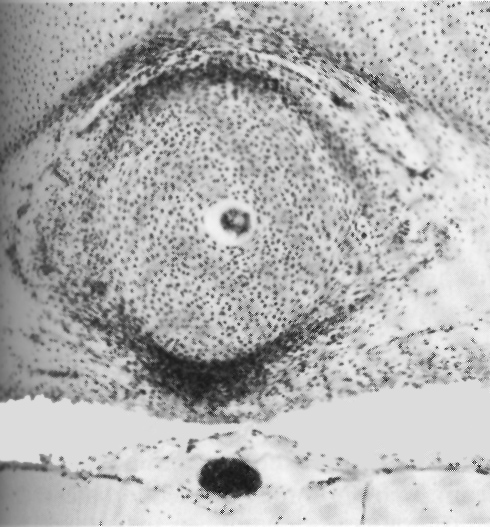
Median atlanto-axial joint at 31 mm, possibly showing the beginning of cavitation.
O'Rahilly, Müller and Meyer (1983)
Fig. 7b
O'Rahilly, Müller and
Meyer (1983) Fig. 7c
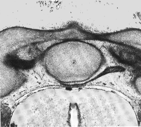

Transverse ligament of atlas at 31 mm.
Dura at stage 23. A. The developing falx is still predominantly leptomeningeal. B. A large cisterna is visible between the basal part of the right cerebral hemisphere and the dural limiting layer.
.
Dural limiting layer,
pachymeninx and skeleton at stage 23.
O'Rahilly, Müller and Meyer (1983) Fig 7e
O'Rahilly and Müller (1986) Fig. 6
O'Rahilly and Müller (1986) Fig.
7